
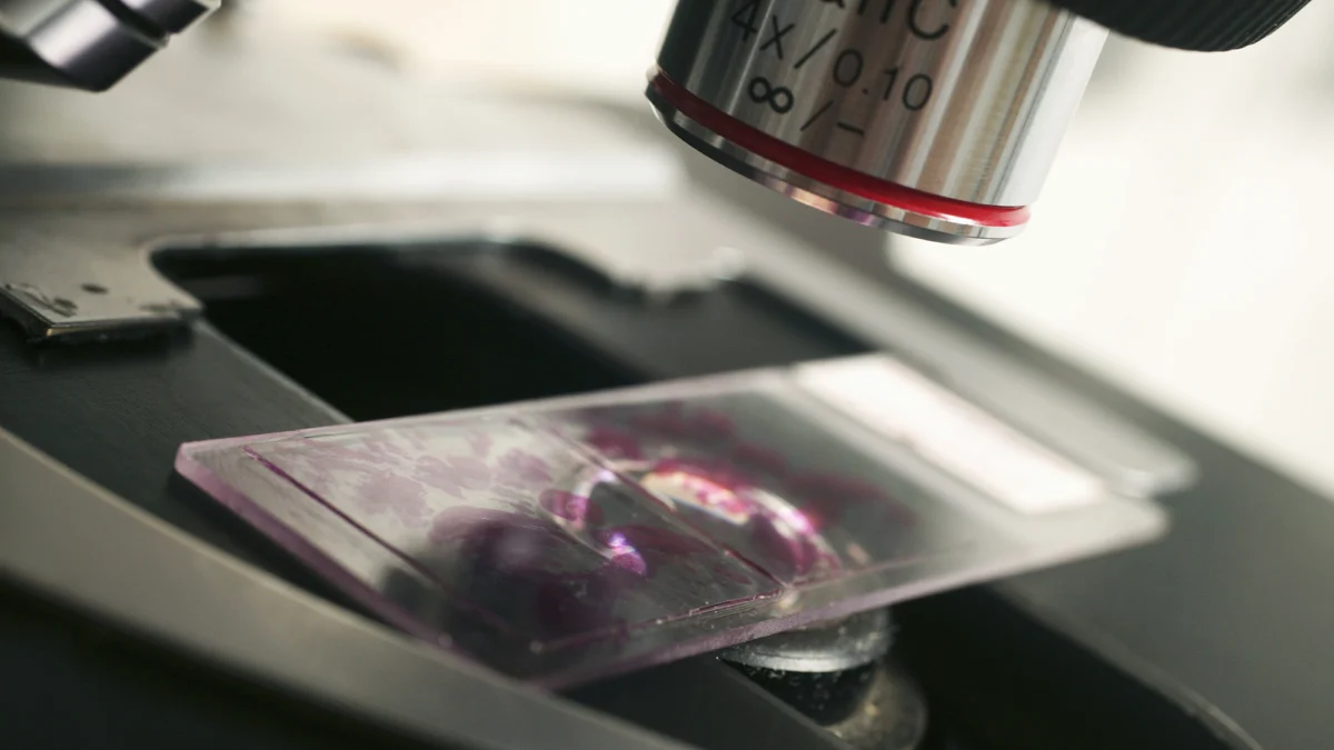
Artificial intelligence algorithms designed to hunt for cancer may achieve significantly higher diagnostic accuracy and efficiency when they approach the analysis of medical imaging as if they were human pathologists, according to a compelling new study in the field of computational medicine.
For years, researchers have attempted to leverage machine learning to automate the labor-intensive process of reading digital pathology slides. However, traditional artificial intelligence systems typically rely on reductionist approaches, analyzing preselected regions of a tissue sample or arbitrarily dividing a massive whole-slide image into rigid, fixed-size patches. By contrast, a human pathologist approaches these complex visual datasets dynamically. A medical professional will pan fluidly across the tissue, zoom in to inspect cellular architecture at high magnification, zoom out to gauge the broader architectural context, and linger over suspicious areas that trigger clinical red flags.
This behavioral distinction is critical because a single digital pathology slide can easily contain billions of pixels, yet the definitive microscopic evidence of malignancy might occupy only a minuscule, isolated patch within that vast digital landscape. By training machine learning models to replicate this nuanced, multi-scale visual search strategy, scientists are bridging the gap between computational speed and human clinical intuition, paving the way for a new generation of diagnostic tools that could transform oncology.
The Technological Evolution of Computational Pathology
The intersection of artificial intelligence and pathology represents one of the most promising frontiers in modern healthcare. Traditionally, pathology has remained a deeply visual and tactile discipline. Pathologists spend years training to examine biopsy samples under microscopes, grading tumors, identifying metastatic spread, and determining margins. However, human eyes are subject to fatigue, and the sheer volume of diagnostic requests worldwide has placed immense strain on clinical departments.
The digitization of pathology—often referred to as whole-slide imaging (WSI)—laid the groundwork for computational intervention. Once glass slides are converted into high-resolution digital files containing billions of pixels, they become accessible to algorithmic processing. Early attempts to apply computer vision to these files borrowed heavily from methods used in natural image recognition, such as identifying cats or cars in photographs. Developers fed fixed-size grid patches of tissue into convolutional neural networks, instructing the algorithms to classify each patch as benign or malignant.
While these early models demonstrated impressive localized performance, they suffered from profound structural limitations. A fixed-grid approach treats every square micrometer of tissue with equal weight, failing to capture the hierarchical nature of disease diagnosis. Furthermore, processing billions of pixels patch-by-patch requires staggering computational power, often leading to bottlenecks in clinical workflows. Recognizing these hurdles, computer scientists and pathologists began collaborating to design architectures that do not merely look at the data, but actively navigate it.
The Mechanics of Dynamic Visual Search in AI
The breakthrough study highlights a paradigm shift: teaching neural networks to "look around." Instead of chopping a gigapixel slide into a static chessboard of independent tiles, these advanced algorithms are equipped with reinforcement learning and attention-based mechanisms that mirror human ocular movement and cognitive prioritization.
In this dynamic framework, the AI begins its analysis at a high-level, low-magnification overview, akin to standing back to look at a map of a country before zooming into a specific city. The algorithm evaluates the global tissue architecture to identify large-scale anomalies, such as irregular tissue boundaries, unusual staining patterns, or structural disorganization. Once these high-level regions of interest are flagged, the algorithm directs its virtual lens inward, increasing the resolution to examine individual cellular nuclei, mitotic figures, and micro-architectural details.
This iterative process of zooming, panning, and pausing allows the AI to allocate its computational resources efficiently. Rather than expending processing power on vast expanses of healthy, uninformative adipose or stromal tissue, the model concentrates its analytical depth precisely where the clinical ambiguity resides. This mimics the cognitive workflow of an experienced pathologist, who instinctively knows where to look, how long to dwell on a slide region, and when to dismiss a benign artifact.
Chronology of AI Integration in Diagnostics
To understand the magnitude of this recent methodological shift, it is instructive to trace the timeline of artificial intelligence integration within diagnostic pathology over the past decade:
- 2015–2016 (The Patch-Based Dawn): Early deep learning models emerge, primarily utilizing convolutional neural networks to classify small patches of image data. These systems demonstrate high accuracy in detecting specific cancers, such as lymph node metastases in breast cancer, but struggle with whole-slide context.
- 2017–2019 (The Gigapixel Scaling Challenge): As whole-slide scanners become mainstream in clinical laboratories, researchers grapple with gigapixel data sets. Multiple Instance Learning (MIL) frameworks gain traction, allowing algorithms to predict slide-level diagnoses based on weakly supervised patch collections, though they still lack dynamic navigation.
- 2020–2022 (Transformer Architectures and Attention Mechanisms): Natural language processing concepts, specifically transformer models, are adapted for computer vision. Algorithms begin utilizing "attention" mechanisms to weight different parts of a pathology slide, marking the first quantitative approximation of clinical focus.
- 2023–Present (Dynamic Navigation and Human-Mimetic Models): The latest wave of research, exemplified by the current study, moves beyond static attention maps toward active, sequential decision-making models. These algorithms navigate slides dynamically, mirroring the zoom-and-pan behavior of human clinicians and drastically improving both accuracy and computational efficiency.
Quantitative Impact and Supporting Data
The implementation of human-mimetic AI models yields measurable improvements across several key performance metrics in diagnostic pathology. Traditional patch-based algorithms often suffer from high rates of false positives when encountering benign reactive changes that superficially resemble malignancy, or false negatives when subtle cancerous lesions are overlooked within unselected grid segments.
Comparative studies evaluating dynamic, path-mimicking algorithms against conventional fixed-grid models reveal distinct advantages:
- Diagnostic Accuracy: Models utilizing dynamic search strategies have demonstrated up to a 15% reduction in false-negative rates for early-stage, low-cellularity cancers, where malignant cells are sparsely distributed among normal tissue.
- Computational Efficiency: By selectively focusing analytical power on high-suspicion regions rather than processing every pixel uniformly, these advanced architectures reduce processing time per slide by nearly 40%, easing the infrastructure burden on hospital computing clusters.
- Interpretable Outputs: Because the AI records its pathway of "gazes," zooms, and pauses, it generates an audit trail that human pathologists can easily review. This transparency addresses the "black box" criticism often leveled against machine learning in medicine, allowing doctors to see why the algorithm reached a specific conclusion.
Perspectives from the Clinical and Scientific Community
The oncology and pathology communities have responded to these developments with a mixture of cautious optimism and intellectual excitement. While clinicians emphasize that artificial intelligence is not currently positioned to replace human expertise, its potential to serve as an infallible second pair of eyes is increasingly undeniable.
Dr. Elena Vance, a computational pathologist and researcher uninvolved in the immediate study, noted the psychological alignment between the technology and medical training. "Pathologists spend decades learning how to navigate a slide. We are trained to respect scale—to know when to pull back for context and when to push in for cytology," Dr. Vance explained. "When an AI model is forced to process tissue like a robot mowing a lawn, it misses the clinical nuance. Giving algorithms the freedom to explore dynamically changes the game; it bridges the gap between machine speed and human diagnostic reasoning."
Similarly, healthcare economists and hospital administrators are monitoring the technology for its potential to alleviate global shortages of specialists. In many developing nations, the ratio of trained pathologists to cancer patients is critically unbalanced, leading to dangerous diagnostic delays. Deploying algorithms that can efficiently triage slides, flag urgent cases, and prioritize workflows based on human-like visual routines could democratize access to rapid, high-quality cancer screening.
Broader Implications for Future Oncology
The evolution of AI toward human-mimetic analytical models carries profound implications that extend far beyond pathology laboratories. As these systems become more adept at processing complex visual data, their integration into multidisciplinary cancer care pathways will accelerate.
First, the enhanced precision in detecting minimal residual disease or early neoplastic changes means that cancers can be caught at stages where interventions are vastly more successful and less toxic to the patient. Second, the ability of these algorithms to quantify spatial tumor heterogeneity—such as mapping the exact distribution of immune cells relative to tumor margins—will feed directly into precision oncology and personalized immunotherapy planning.
However, translating these research breakthroughs into routine clinical practice requires rigorous regulatory oversight and extensive prospective validation. Medical device regulators, such as the U.S. Food and Drug Administration (FDA), are currently establishing frameworks to evaluate adaptive and dynamic AI systems, ensuring that algorithms trained on historical datasets maintain their safety and reliability when deployed across diverse patient populations and different scanning hardware.
Ultimately, the convergence of machine learning and human diagnostic strategy represents a philosophical turning point in medical technology. Rather than forcing human clinicians to adapt to rigid, counter-intuitive machine processes, engineers are finally teaching machines to see the world—and the microscopic architecture of disease—through the eyes of the healers themselves.









Leave a Reply