
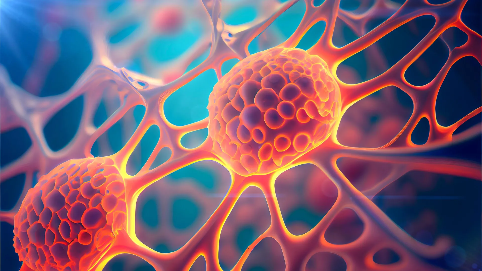
For over half a century, the biological mystery of how damaged epithelial tissues orchestrate massive, coordinated regeneration has remained a central enigma in developmental and cell biology. While scientists have long observed the phenomenon known as "compensatory proliferation"—the process by which organisms replace destroyed cells with fresh, functional tissue—the underlying molecular architecture triggering this recovery has eluded researchers. Now, a landmark study conducted by the Weizmann Institute of Science, published in the journal Nature Communications, has identified a sophisticated survival mechanism that explains how cells navigate the brink of destruction to rebuild damaged organs. This discovery not only sheds light on the resilience of skin and epithelial linings but also offers a troubling insight into why certain cancers exhibit lethal resistance to radiation therapy and return with increased aggression.
A Historical Perspective on Compensatory Proliferation
The scientific foundation for this discovery dates back to the 1970s. During that era, pioneering researchers exposed fruit fly (Drosophila) larvae to high doses of ionizing radiation. Despite the widespread cellular trauma caused by the radiation, the larvae demonstrated an extraordinary capacity to regenerate fully functional wings. This observation established that epithelial tissues possess an inherent "self-repair" program capable of being activated following catastrophic injury. Over the subsequent decades, similar regenerative phenomena were documented across various species, including mammals, suggesting that this mechanism is a fundamental, evolutionary conserved response to injury. However, the specific molecular "switch" that distinguishes a cell destined for death from one that will lead the charge in regeneration remained unknown until the recent work led by Professor Eli Arama of the Weizmann Institute’s Molecular Genetics Department.
The Duality of Caspases: From Destruction to Regeneration
At the heart of the cellular life cycle is apoptosis, a highly regulated form of programmed cell death. When a cell sustains irreparable damage, a cascade of enzymes known as caspases is activated. Traditionally, these enzymes are categorized as the "executioners" of the cell; an initiator caspase triggers the process, and effector caspases follow, systematically dismantling internal proteins to clear the way for new, healthy cells.
For the past twenty years, however, researchers have observed that caspases do not always follow the path to destruction. Prof. Arama and his colleagues hypothesized that these enzymes might possess non-lethal, secondary functions that could explain compensatory proliferation. To test this, a team led by Dr. Tslil Braun utilized advanced genetic tracing tools to monitor the behavior of epithelial cells in fruit flies exposed to ionizing radiation. By employing a delayed sensor, the team identified a unique population of cells that initiated the self-destruct sequence but, instead of perishing, survived and began to proliferate. The team designated these cells as "DARE" cells (Death-Associated Recovery Cells).
The Mechanism of DARE and NARE Cells
The research revealed that DARE cells are pivotal to the repair process. Within 48 hours of irradiation, these cells accounted for nearly 50% of the replenished tissue. When the researchers experimentally depleted DARE cells, the entire compensatory proliferation response collapsed, proving their necessity for successful regeneration.
The study further identified a second, distinct population termed "NARE" cells (Non-death-Associated Recovery Cells). Unlike their DARE counterparts, NARE cells never activated the initiator caspase. The researchers discovered a complex, symbiotic feedback loop between these two groups: DARE cells secrete growth factors that stimulate NARE cells, while NARE cells produce inhibitory signals that prevent the DARE population from over-proliferating. This delicate, homeostatic crosstalk ensures that tissue regeneration is precise, preventing the runaway growth that characterizes tumor formation.
Why DARE Cells Survive: The Role of Molecular Motors
A critical aspect of the study was determining how DARE cells evade their death sentence. The team discovered that the apoptotic pathway in DARE cells stalls at the initiator stage, preventing the executioner caspases from completing the destruction. This stall is mediated by a specific "molecular motor" protein. This protein anchors the initiator caspase to the cell membrane, physically sequestering it from the targets it would otherwise destroy.
This finding carries significant implications for oncology. The research team noted that the overactivation of this specific motor protein has been previously observed in human cancerous tumors. If cancer cells hijack this natural survival mechanism, they effectively render themselves "death-proof" against standard therapies like radiation, which rely on inducing apoptosis to eliminate tumor mass.
Clinical Implications and Future Directions
The potential for this discovery to reshape cancer treatment protocols is substantial. Data from the experiment indicated that the descendants of DARE cells are not merely survivors; they are biologically hardened. When the tissue was subjected to a second round of radiation, the descendants of DARE cells were found to be seven times more resistant to death than the original cell population. This finding provides a compelling explanation for the clinical observation that recurrent tumors are often more aggressive and resistant to follow-up treatments than primary tumors.
"Many cancers originate in epithelial cells that have lost normal growth control," explains Prof. Arama. "Many traditional cancer treatments aim to cause them to self-destruct through apoptosis. Our findings pave the way for understanding why such treatments sometimes fail and how they could be improved."
Broader Scientific Analysis
The work of the Weizmann team, which included collaboration with Prof. Andreas Bergmann of UMass Chan Medical School and Prof. Luis Alberto Baena-Lopez of the Severo Ochoa Molecular Biology Center, highlights a fundamental paradox in regenerative medicine: the same mechanisms that allow the body to heal itself from injury can be subverted to facilitate disease.
The immediate next steps for the research community involve translating these findings from fruit fly models to human biological systems. If the same caspase-stalling mechanism exists in humans, it may eventually be possible to develop adjuvant therapies that "unlock" the apoptotic pathways in tumor cells, making them susceptible to radiation once again. Conversely, for conditions involving tissue loss—such as chronic wounds or organ damage—researchers might one day be able to safely stimulate the DARE-cell mechanism to accelerate natural healing.
This discovery marks a transition in the understanding of cellular biology from a binary view of "life or death" to a more nuanced appreciation of the gray areas in between. By identifying the specific molecular motors and signaling pathways involved, scientists have moved closer to a future where medical intervention can specifically modulate these pathways to either enhance repair or suppress the persistent survival of malignant cells. As the study moves toward clinical validation, it remains a testament to the power of fundamental research in elucidating the core mechanisms that define human health and pathology.








Leave a Reply